| Vascular Structure and Function |
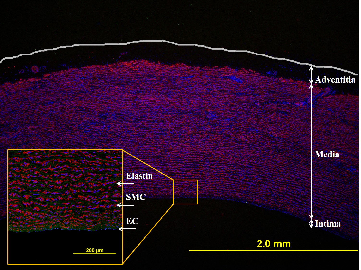 |
We seek to better understand the interactions between cells and their environment. The image on the left is cross-section of a porcine aorta. This aorta is comprised of 3 layers. The outmost layer is called the adventitia. The middle layer the media and the layer closest to the blood stream, the intima. The media is comprised of multiple layers of alternating elastin and smooth muscle cells. The elastin give the artery the ability to "recoil" after each beat of the heart and surge of blood. The intima is comprised of a monolayer of endothelial cells (ECs). These cells are important for the acute vasoactivity of the artery. That is they respond to changes in flow by releasing molecules depending on the situation. For example, if you need more blood flow, your heart will beat faster, causing a build up of fluid inside the artery. Well the ECs will release nitric oxide (NO) a vasodilator that will expand the inner diameter and allow the fluid to get to where it needs to go. |
|
| Cell Biophysics and Mechanobiology |
 |
Investigating biophysical and mechanobiological aspects of leukocyte trans-endothelial migration. |
|
|
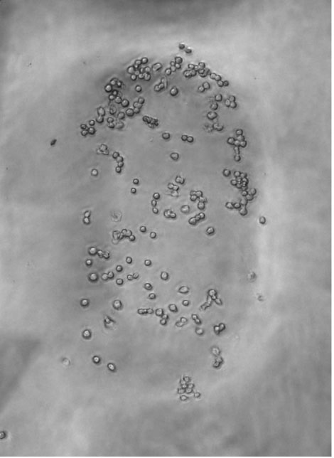 |
Testing how cells will respond to their mechanical environment. After 48 hrs. macrophages are preferentially attached to the stiffer areas of the hydrogel.Pubmed Link |
|
|
|
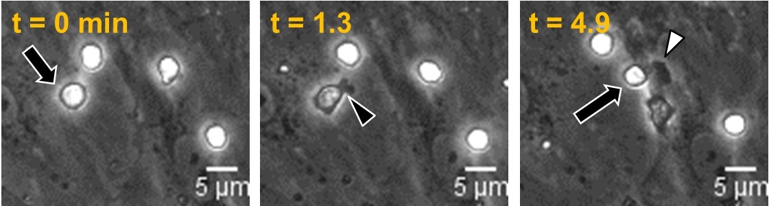
Interactions between cells as a function of stiffness. This video shows monocytes (white cells) migrating on the an human aortic endothelial monolayer (gray cells). The cell proceeds to transmigrate between two endothelial cells. The trans-endothelial migration process takes about 2 minutes when the endothelial cells are on stiff substrates(>5 kPa) and about 4 minutes on softer (1 kPa) substrates. The movie playback is 50x faster than reality. |
|
| Multiscale Modeling |
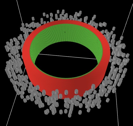 |
We use computer modeling to predict how arteries will respond to their environment over time. For example, agent based models allow cells to be treated as nearly autonomous agents that react to their environment according to a set of "rules" or functions. The artery segment shown on the left is a representation of a human left anterior descending artery (LAD). The 3 main cell types (endothelial, smooth muscle and fibroblast) are shown in different colors. |
|
|
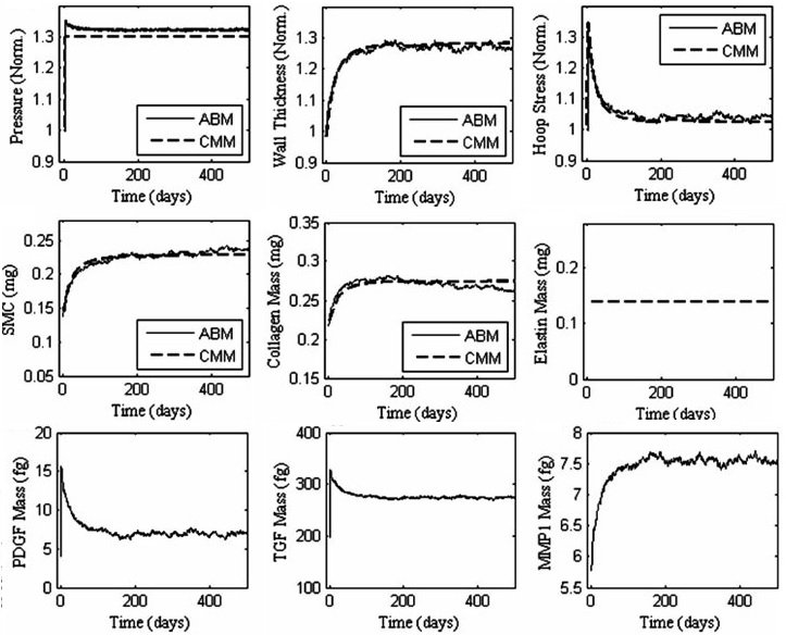 |
Predicting the geometry, mechanics and soluble factors as an artery remodels due to mechancial or chemical stimuli.rent colors. This example simulates a 30% increase in blood pressure after 42 hrs. This pertubation results in an increase in hoop stress (i.e. circumferential stress) and a gradual accumulation of collagen and smooth muscle. In addition we see a gradual increase in metalloproteinases. Moreover growth factors initially increase and then gradually decrease. This integrative process between mechanics and biological milieu occurs in attempt to restore the homeostatic value of hoop stress in the arterial wall. Pubmed Link |
|